Induced change microscopy of live cells
Induced Change Microscopy of Live Cells

My PI just informed me our new project is to investigate the instantaneous effect of a compound on live cells. Some of the setup is intuitive but now I have run into issues I didn’t realize would be problematic. For instance in order to observe my cells what kind of containment is best to use? How do I expose the cells to an alternate media without compromising them? How do I precondition my media? How long does it take to acquire the expected changes? Who can help?
Research Fellow at Stanford University
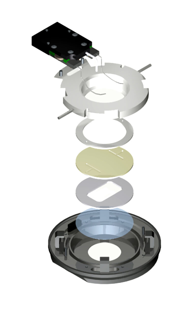
Fortunately, you are not the first person to encounter these issues. As it turns out, this is a routine process used to investigate many cellular reactions. One by one in order, the type of containment vessel should be a parallel plate flow cell having a coverslip surface for imaging. There are several commercial ones available. The one most recommended is the Bioptechs FCS2.
When comparing to the others, the FCS2 was the only one that allows you to select or create any flow geometry you want including laminar flow instead of being rigidly confined to the manufacturers design. It also provided temperature support uniformly over the entire aperture of the chamber. It is compatible with all modes of microscopy you may intend to use and is easy to assemble. When it comes to perfusion you may be thinking it is a plumber’s nightmare but in fact it is already worked out. If you are working with two sources of media, one is just a nutrient media and the other has the variant factor. The plan is to image cells on the scope for about 30 minutes with nutrient media to demonstrate they are “happy” and not compromised in their foreign environment. Then, depending on experimental protocol, introduce the alternate media containing your variant factor while acquiring images of the effects.
If the expected reaction takes place slowly you can manually introduce the variant, but if the protocol requires a rapid introduction of the variant that is where it gets a little tricky and is best to automate the process. There are two sources of media coming together external to the optical cavity where the cells are plated. Therefore, there is a dead-volume between the adjoining flows and the cells, and a small diffusion gradient that occurs when one flow follows another. The goal is to get the variant factor to the cells at its source concentration ASAP to record the immediate reaction.
To accomplish this therecomputer programs that control the flow rate of two independent, smooth flow, peristaltic perfusion pumps.
The program allows you to; establish a maintenance flow of nutrient media for a predetermined amount of time that you control, then ramp down the nutrient flow while accelerating the flow of the variant factor, then sustain that rate for a for another determined period of time you chose to ensure the concentration of the variant media is at its peak. The Variant flow rate is then de-accelerated to a minimum rate for imaging. The process can then repeated to rinse the variant factor and restore the prior environmental conditions. This saves a lot of headache
making these transitions especially when it has to be consistently repeated many times for statistical reasons
If you can’t get away with hepes media, you will need to sustain a CO2 tension in the media to maintain proper pH. A number of CO2 systems are available but the one that makes the most sense starts with a 5% CO2 and air tank under pressure then reduces the pressure to ambient with a regulator. That way the gas mixture can be metered out by virtue of its volume with a simple peristaltic pump. If you place a 14 gauge tube into a flask containing media each bubble that rises is about 15 micro-liter. Therefore, all you have to do to establish the flow rate is count the bubbles per minute and do a little arithmetic!
The amount of time it takes to acquire results will depend on the concentration of the variant factor and your cells.